Non-Ossifying Fibroma (NOF)
Non-ossifying fibroma (NOF) is a benign, non-osteogenic bone lesion composed of fibroblastic cells, typically located in the metaphysis of long bones during childhood and adolescence.
It is usually asymptomatic and detected incidentally on radiographs obtained for other reasons.
1.Synonyms
Fibrous cortical defect (for smaller lesions)
Metaphyseal fibrous defect
Fibroxanthoma
Nonosteogenic fibroma
Fibrous histiocytoma of bone
Sometimes confused with “cortical desmoid” (different localization)
2. Associated Conditions / Syndromes
NOF can be an isolated finding but may also be associated with certain systemic syndromes:
Neurofibromatosis type 1 (NF1)
Jaffe-Campanacci syndrome: Multiple NOFs + café-au-lait macules + mental retardation + hypogonadism + cardiac anomalies
3. Epidemiology
Age: Common between 5–20 years
Sex: Slight male predominance (~1.6:1)
Prevalence: Seen radiographically in 30–40% of children
Most frequent locations: Distal femur, proximal tibia, distal tibia, proximal fibula
4. Pathogenesis and Genetics
Previously thought to be a reactive process; current DNA analyses have revealed KRAS, FGFR1, and NF1mutations → suggesting a neoplastic process related to RAS-MAPK pathway activation.
Typically eccentric in location, adjacent to the cortex.
5. Clinical Features
Usually asymptomatic
Large lesions:
Mechanical weakness → risk of pathological fracture
May cause pain
Diagnosis is often facilitated if a pathological fracture is present
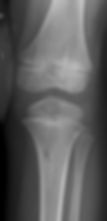
6. Imaging
Radiograph: Eccentric, metaphyseal, cortically based, well-defined, lobulated radiolucent lesion with internal septations
Often 1–3 cm (fibrous cortical defect) or larger (NOF)
CT: Demonstrates cortical thinning and intracortical location
MRI: Hyperintense on T2; hypointense on T1 if hemosiderin present
Periosteal reaction is typically absent
7. Histology
Gross:
Well-circumscribed, soft, yellow-brown fibrous tissue
Cut surface may show small foci of hemorrhage and hemosiderin deposits
Microscopic:
Well-vascularized fibrous stroma with haphazardly arranged spindle-shaped fibroblasts
Interspersed lipid-laden foam cells (xanthomatous histiocytes)
Focal multinucleated giant cells
Frequent hemosiderin pigment deposition
Minimal cellular atypia, rare mitotic figures
Occasional ossification or bone trabeculae within fibrous stroma
Sclerotic bony rim may be present at the periphery
Differential Diagnosis: Aneurysmal bone cyst, fibrous dysplasia, giant cell tumor
8. Treatment and Natural History
Small/asymptomatic: Observation (most regress and sclerose spontaneously by late adolescence)
Large/high fracture risk: Curettage + bone grafting
Prophylactic fixation may be considered
Recurrence after surgery is rare