The shoulder joint is the structure that connects the upper extremity to the axial skeleton and primarily enables the positioning of the hand in 3D space. It is a complex structure formed by the combination and harmonious functioning of 4 different joints:
· Glenohumeral joint
· Acromioclavicular joint
· Sternoclavicular joint
· Scapulothoracic joint
Osseous anatomy of the shoulder girdle
Humerus:
· The average diameter of humeral head is 43mm, with approximate retroversion of 20° from transepicondylar axis of the distal humerus.
· Articular surface has a 130° of superior inclination from the humeral shaft
· Proximal humerus serves as attachment sites for rotator cuff tendons with greater and lesser tuberosities (subscabularis – lesser tuberosity, supraspinatus/infraspinatus/teres minor – greater tuberosity).
Primary blood supply of the humeral head is provided by posterior humeral circumflex artery. Ascending branch of anterior humeral circumflex artery and arcuate artery are other blood sources to the humeral head. Ascending branch of the anterior humeral circumflex artery runs parallel to the lateral aspect of the bicipital groove. Caution should be paid not to damage during plating proximal humerus fractures.
Glenoid has a pear-shaped articular surface that is wider inferiorluy and narrower superiorly. There is an average superior inclination of 5° of glenoid articular surface. Glenoid articular surface can have a version in relation to scapular body ranging from 7° of retroversion to 10° of anteversion (average is 5° of retroversion).
Coracoid is the attachment site for
· Conjoint tendon (coracobrachialis + short head of the biceps)
· Pectoralis minor tendon
· Coracoacromial ligament
· Coracoclavicular ligaments (conoid and trapezoid)
Coracoid serves as an important surgical landmark for deltopectoral approach. Medial to coracoid is home to important neurovascular structures (brachial plexus, axillary artery).
Acromion has 3 secondary ossification centers:
· Meta (basi) acromion
· Meso (mid) acromion
· Preacromion
Failure of fusion of these ossification centers causes “os acromiale”.
Acromion morphology is traditionally classified using Bigliani’s classification, in terms of morphlogical effect on subacromial impingement:
· Type 1: flat
· Type II: curved
· Type III: hooked
Type III acromion is traditionally considered highly associated with subacromial impingement and degenerative rotator cuff tears.
The distance between acromion and humerus is called “acromiohumeral interval”. AHI<6mm is highly associated with subacromial impingement.
Glenohumeral Stability
Stability is primarily achieved by static and dynamic restraints.
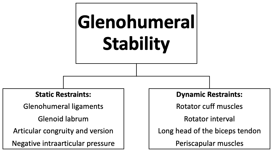
Glenohumeral ligaments:
Glenohumeral ligamets are superior (SGHL), middle (MGHL) and inferior (IGHL) glenohumeral ligaments. These ligaments are among the static restraints to glenohumeral stability
SGHL extends from anterosuperior labrum to humerus. It contributes to formation of biceps pulley and prevents anteroinferior translation of long head of biceps. It serves as a restraint to inferior translation of humeral head at 0° of abduction.
MGHL prevents anterior and posterior translation of humeral head in the midrange of abduction in external rotation.
IGHL is composed of anterior, posterior and superior bands. Posterior band of IGHL is the most important restraint to posterior humeral head subluxation at 90° of flexion and internal rotation. Thightness of posterior band of IGHL causes internal impingement and increases shear forces on superior labrum (associated with SLAP lesions).
Anterior band of IGHL is the priamry restraint to antero-inferior translation of humeral head in 90° of abduction and maximum external rotation. Bankart lesions are injury of to anteroinferior labrum and attachment of inferior band of IGHL to labrum.
Glenoid labrum:
Glenoid labrum is composed of fibrocartilagenous tissues and serves to promote cavity-compression effect of glenuhumeral joint by increasing glenoid socket depth.
Vascular supply to the labrum is not provided from underlying bone. Receinves blood supply fom capsule and periosteal vessels. Anterior-superior labrum has the poorest blood supply (SLAP lesions.)
Anterior band of the IGHL anchors to anterior labrum (weak link causes Bankart lesion). Long head of the biceps tendon anchors to superior labrum (weak link causes SLAP lesion).
Rotator interval:
Rotator interval is an anatomical structure formed by:
· Coracoid base (lateral border)
· Supraspinatus tendon (superior border)
· Subscapularis tendon (inferior border)
· Transvers humeral ligament (lateral border)
Contents of rotator interval:
· Glenohumeral joint capsule
· SGHL
· Coracohumeral ligaments
· Long head of the biceps tendon
Long head of the biceps tendon acts as a dynamic joint restraint by serving as a humeral head depressor. SGHL and subscapularis tendon forms biceps pulley and stabilizes long head of the biceps tendon in the bicipital groove.
Acromioclavicular joint
Acromioclavicular (AC) joint is a diarthrodial joint and contains a fibrocartilaginous intraarticular disc. The motion of the AC joint is primariliy limited to gliding motion. Its contribution to rotation is minimal.
The stability of AC joint is provided by acromioclavicular ligaments, coracoclavicular ligaments, deltotrapezial fascia, deltoid and trapezius
Acromioclavicular ligament
· Superior, inferior, anterior and posterior components. Most important ones are superior and posterior (critical for stability following distal clavicle resection)
· Mainly supports horizontal stability
Coracoclavicular ligaments
· Composed of two ligaments (conoid and trapezoid)
· Conoid is medially (approximately 4.5 cm from lateral clavicle end) and trapezoid is laterally ( 3cm from lateral clavicle end) located.
· Important for vertical stability
Sternoclavicular joint
Sternoclavicular (SC) joint is a diarthrodial (like AC joint) saddle joint. The joint surfaces are incongrous with approximately 50% are contact between sternum and clavicle. The joint contains a fibrocartilaginous intraarticular disc.
The stability of SC joint is secured by:
· Posterior sternoclavicular ligament
· Anterior sternoclavicular ligament
· Costoclavicular (rhomboid) ligament
· Intraarticular disk ligament
Clavicle is the first bone to start ossification and is the last bone to complete. Ossification process of the clavicle start at 5-6th gestation week and ends at around 20-25 years (medial epiphysis of the clavicle). Most injuries of SC joint before 25 years of age are medial physeal fractures of SC joint, rather than SC joint dislocations.
Scapulothoracic Joint
Scapulothoracic (ST) joint is an articulation between scapula and thorax, rather than a true joint. The scapula is
· 10-20° anteverted
· 30-45° internally rotated
· 3° superiorly inclined in resting position.
One third of total shoulder abduction is performed by the motion of the ST joint (First 120° by glenohumeral joint, last 60° by ST joint)