Overview
● A Chance fracture is an unstable spine fracture primarily occurring at the thoracolumbar junction (T10-L2).
● It is characterized by a horizontal fracture line extending from posterior to anterior through the spinous process, pedicles, and vertebral body.
● The injury results from flexion-distraction forces, typically due to rapid deceleration in motor vehicle collisions where the lap belt acts as a fulcrum.
● These fractures often occur in children and young adults, with a male predominance.
Pathophysiology
● According to Denis's three-column concept, the injury involves distraction of the middle and posterior columns, rendering it unstable.
● Classification:
○ Osseous: Involves fractures of the spinous process, pedicles, and vertebral body.
○ Ligamentous: Involves rupture of the interspinous ligament, ligamentum flavum, facet capsule, and potentially the posterior longitudinal ligament.
Clinical Presentation
● Patients rarely present with neurological deficits unless there is retropulsion of bone fragments causing cord or cauda equina contusion.
● "Seatbelt Sign": Bruising or abrasion across the abdomen is a significant physical finding associated with Chance fractures.
● Associated Injuries: The incidence of concurrent intra-abdominal injuries (e.g., bowel perforation, mesenteric laceration) can be as high as 50%.
Imaging
Computed Tomography (CT): This is the most appropriate initial imaging for trauma patients and is superior to radiography for fracture detection. It can also detect the "seatbelt sign" as stranding in the subcutaneous fat of the abdominal wall.
Magnetic Resonance Imaging (MRI): MRI is superior to CT for detecting soft tissue injuries (ligaments, spinal cord, intervertebral discs) and is indicated if
clinical findings suggest ligamentous injury.
Radiography: Plain films are less sensitive; findings may include distraction of spinous processes or facet joints.
Classification
The AO Spine Thoracolumbar Injury Classification System is the current standard, categorizing Chance fractures as Type B (tension band) injuries.
● Type B1 (Bony Chance Fracture): Monosegmental, purely osseous failure. The fracture line extends through the spinous process, pedicles, and vertebral body, with the PLC remaining intact or avulsed with bone.
● Type B2 (Osseoligamentous Injury): Disruption of the posterior tension band ligaments with or without associated bony injury. These injuries are inherently unstable and have poor healing potential without surgical stabilization.
● Type C Injury: Flexion–distraction injury with superimposed translation or displacement, representing a highly unstable pattern.
Treatment
Conservative Management:
● Appropriate for patients with purely osseous injuries and no neurological deficits.
● Immobilization in a cast or Thoracolumbosacral Orthosis (TLSO) with hyperextension for 8-12 weeks.
● Outcome: Bony healing generally occurs with time, and union rates are high.
Surgical Management:
● Indicated for ligamentous injuries, neurological deficits, or patients with obesity where orthosis is not feasible.
● The predominant method is long-segment posterior fixation using pedicle screws, with or without interbody fusion.
● Percutaneous techniques may reduce morbidity compared to open surgery.
Prognosis & Complications
● Outcomes are generally good; surgical stabilization yields >90% good results after one year.
● Complications: The most common long-term complaint is residual low back pain. Residual kyphosis may also occur.
● Morbidity: High morbidity is often associated with delayed diagnosis of concomitant intra-abdominal injuries rather than the fracture itself.
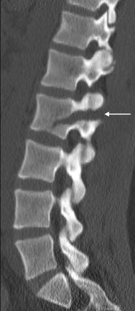
Dissolving pedicle” sign in 28-year-old woman. Sagittal CT reformation depicts horizontal L2 Chance fracture through left pedicle and into vertebral body (arrow).
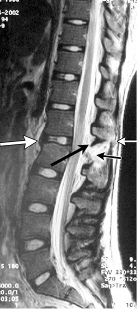
Soft-tissue injuries. Sagittal T2-weighted: Small white arrow: Torn supraspinous ligament (part of the Posterior Ligamentous Complex disruption). Small black arrow: Torn interspinous ligament (part of the Posterior Ligamentous Complex disruption). Large black arrow: Torn ligamentum flavum (part of the Posterior Ligamentous Complex disruption).Large white arrow: Buckling of the anterior longitudinal ligament.
Reference
1. Durel R, Rudman E, Milburn J. Clinical images - a quarterly column: chance fracture of the lumbar spine. Ochsner J. 2014 Spring;14(1):9-11. PMID: 24688326; PMCID: PMC3963060.
2. Elhadad, L.; Mathews, S.; Toma, J.; Gulati, A.; Masri, D. Chance Fracture: A Case Report and Review of the Literature. Radiology Case Reports 2025, 20 (1), 680–685. https://doi.org/10.1016/j.radcr.2024.10.074.
3. Serag I, Scherer J, Popescu EC, Costachescu B, Holas M, Alahmadi AS, Aly MM. A meta-analysis of the incidence of intra-abdominal injuries associated with thoracic or lumbar flexion-distraction injuries. Injury. 2025 Jun;56(6):112337. doi: 10.1016/j.injury.2025.112337. Epub 2025 Apr 8. PMID: 40273660.
4. Vaccaro AR, et al. "AO Spine Thoracolumbar Classification System." Spine, 2020 update
5. Bernstein MP, Mirvis SE, Shanmuganathan K. Chance-type fractures of the thoracolumbar spine: imaging analysis in 53 patients. AJR Am J Roentgenol. 2006 Oct;187(4):859-68. doi: 10.2214/AJR.05.0145. PMID: 16985126.
6. Groves, C. J.; Cassar-Pullicino, V. N.; Tins, B. J.; Tyrrell, P. N. M.; McCall, I. W. Chance-Type Flexion-Distraction Injuries in the Thoracolumbar Spine: MR Imaging Characteristics. Radiology 2005, 236 (2), 601–608. https://doi.org/10.1148/radiol.2362040281.