Classification of Failure
A. Oncological Failure
1. Local Recurrence
Intralesional surgery
Contaminated surgical field
Inadequate surgical margins
Skip lesions
Incorrect biopsy tract placement
2. Systemic Progression
Metastatic disease
Resistance to chemotherapy
Dedifferentiation
3. Misdiagnosis
Errors in distinguishing benign from malignant lesions
Limitations of frozen section analysis
Discordance between pathological and radiological findings
Particular attention should be paid to the following concepts:
Whoops surgery: The unplanned excision of a musculoskeletal tumor that was not recognized as malignant, performed without adherence to oncological principles and with inappropriate surgical margins.
Unplanned excision: Excision performed without adherence to oncological principles because the lesion was not recognized as a sarcoma. These procedures are generally carried out without appropriate MRI evaluation, without proper biopsy principles, or with inadequate surgical margins. The treatment algorithm is presented in Figure 1.
Contaminated field: Surgical disruption of the natural biological boundaries of the tumor, resulting in tumor spread and contamination of surrounding tissues. The most common causes are improper biopsy techniques and hematoma dissemination.
Failed biopsy: A spectrum of errors occurring during biopsy acquisition or the diagnostic process. This includes tumor seeding, hematoma spread, and contamination of adjacent compartments.
Management in these situations includes the following steps:
Restaging:
Contrast-enhanced MRI, computed tomography (CT) for staging purposes, and positron emission tomography (PET) imaging when indicated.Repeat biopsy:
Recommended when the initial pathology is suspicious, the tumor grade remains uncertain, or there is discordance between radiological and pathological findings.Wide re-excision:
This constitutes the cornerstone of treatment and should be planned meticulously to achieve adequate oncological margins. (Figure 1)
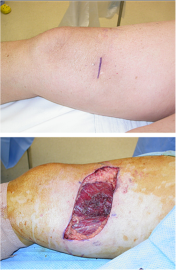
Figure 1 A poorly oriented transverse incisional biopsy for a superficial, high-grade sarcoma of the medial right thigh. Wide resection to incorporate the previous biopsy scar required a generous, transversely oriented excision.
Radiotherapy:
Frequently required because of the high risk of microscopic tumor dissemination and residual contamination.Amputation:
May be necessary in cases of extensive contamination, major neurovascular involvement, joint contamination, or when limb-salvage procedures are unlikely to provide meaningful functional outcomes.
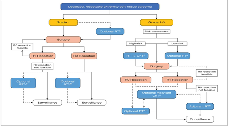
Figure 1: Treatment algorithm recommended by European Society for Medical Oncology (ESMO) after unplanned resection in localized resectable soft tissue tumors
3. Surgical Margin Problems
Types of Surgical Margins
Intralesional
Marginal
Wide
Radical
One of the most important causes of failure includes:
Excessive emphasis on preservation of neurovascular structures at the expense of oncological safety
Dissection through the reactive zone
Failure to excise the biopsy tract during definitive tumor surgery
4. Mechanical Failure
This section is particularly important in the setting of megaprosthetic reconstruction.
Henderson Classification
The Henderson classification remains the most widely used system for classifying failure after limb-salvage reconstruction.
Type 1 — Soft Tissue Failure
Instability
Dislocation
Abductor insufficiency
Loss of the extensor mechanism
Type 2 — Aseptic Loosening
Stem loosening
Stress shielding
Osteolysis
Type 3 — Structural Failure
Stem fracture
Fatigue fracture
Modular junction failure
Type 4 — Infection
One of the most feared complications in orthopedic oncology reconstruction
Type 5 — Tumor Progression
Local recurrence
New metastatic destruction
The Henderson classification is presented table 1.
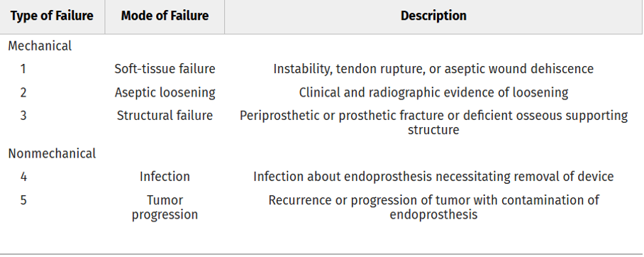
Table 1: Classification of Segmental Endoprosthetic Failure - Henderson Classificaiton
5. Biological Failure
Allograft Failure
Nonunion
Delayed union
Resorption
Fracture
Infection
Devitalized Bone Reconstructions
Autoclaved autograft
Irradiated autograft
Liquid nitrogen-treated autograft
Pasteurization
Common problems associated with these reconstruction methods include:
Slow creeping substitution:
Creeping substitution refers to the gradual biological process in which devitalized bone is resorbed and replaced by newly formed viable bone over time. It represents a fundamental component of biological reconstruction. However, complications such as nonunion, graft fracture, resorption, and infection may occur during this process.Revascularization problems
Osteoporotic bone changes
6. Infection
Infection is a common and challenging complication in orthopedic oncology.
Risk Factors
Prolonged surgical procedures
Large dead space formation
Radiotherapy
Chemotherapy
Immunosuppression
Inadequate soft tissue or muscle coverage
Management
Debridement with implant retention
One-stage revision
Two-stage revision
Silver-coated megaprostheses
Muscle flap reconstruction, including:
Vertical rectus abdominis myocutaneous (VRAM) flap
Gastrocnemius flap
Latissimus dorsi flap
7. Failure in Pelvic Tumor Surgery
Failure rates are particularly high in pelvic tumor surgery because of several factors, including:
Complex pelvic anatomy
Difficulty in achieving adequate surgical margins
Challenges related to load transfer and biomechanics
Formation of large dead spaces
Common Modes of Failure
Collapse of Harrington reconstructions
Cage failure
Screw loosening
Lumbopelvic instability
Wound complications
Particular attention should be given to:
Reconstruction following internal hemipelvectomy
Periacetabular reconstruction
Flail hip conditions
Ice-cream cone prostheses
These reconstruction methods and their associated complications may be discussed in detail.
8. Failure in Spine Tumor Surgery
Mechanical Failure
Rod fracture
Cage subsidence
Junctional kyphosis
Oncological Failure
Epidural tumor progression
Failure of en bloc resection
Biological Failure
Failure of spinal fusion following radiotherapy
Particular emphasis should be placed on:
Separation surgery
Carbon fiber implants
The relationship between stereotactic radiotherapy and spinal reconstruction outcomes
9. Salvage Strategies
Revision Options
Revision megaprosthesis
Total femoral replacement
Three-dimensional (3D) printed implants
Custom-made pelvic prostheses
Biological Salvage Procedures
Vascularized fibular grafts
Allograft–prosthetic composites
Last-Resort Procedures
Amputation
Rotationplasty
Hip transposition
10. Psychological and Functional Failure
This is a frequently overlooked yet highly important aspect of failure in orthopedic oncology.
Key considerations include:
Musculoskeletal Tumor Society (MSTS) score
Toronto Extremity Salvage Score (TESS)
Patient expectations
Chronic pain
Psychological and physical burden associated with repeated surgical procedures
Body image disturbances and impaired self-perception
References:
1) Henderson ER, Groundland JS, Pala E, et al. Failure mode classification for tumor endoprostheses: retrospective review of five institutions and a literature review. J Bone Joint Surg Am. 2011;93(5):418-429. doi:10.2106/JBJS.J.00834
2) Gronchi A, Miah AB, Dei Tos AP, et al. Soft tissue and visceral sarcomas: ESMO–EURACAN–GENTURIS Clinical Practice Guidelines for diagnosis, treatment and follow-up. Ann Oncol. 2021;32(11):1348-1365. doi:10.1016/j.annonc.2021.07.006
3) Fromm J, Klein A, Mentrup F, et al. Unplanned resections of soft tissue sarcomas: necessity of re-resection? Cancers (Basel). 2024;16(10):1851. doi:10.3390/cancers16101851
4) Siegel GW, Biermann JS, editors. Orthopaedic Knowledge Update: Musculoskeletal Tumors 5. 5th ed. Rosemont, IL: American Academy of Orthopaedic Surgeons; 2020.
5) Tedesco NS, Henshaw RM. Unplanned resection of sarcoma. J Am Acad Orthop Surg. 2016;24(3):150-159. doi:10.5435/JAAOS-D-15-00074