Overview
In young patients they follow moderate trauma and are mechanically stable. In older adults they are mostly osteoporotic fragility fractures, the most common osteoporotic fracture, and many are clinically silent. An incident vertebral fracture sharply increases the risk of further fractures; about one in five patients sustains a new vertebral fracture within a year [5]. Every fracture is therefore a sentinel event for osteoporosis.
Clinical Presentation
Osteoporotic fractures cluster in the mid-thoracic spine and at the thoracolumbar junction. Acute axial pain after minimal or no trauma is typical, often worse on standing and relieved by lying down. Neurological deficits are rare unless the posterior wall is involved. Chronic presentations include height loss, progressive thoracic kyphosis, early satiety, reduced pulmonary capacity and, in severe cases, painful impingement of the lower ribs on the iliac crest. Night pain, weight loss, fever or a history of cancer mandate investigation for malignancy or infection.
Imaging
Lateral radiographs establish the diagnosis and allow semiquantitative grading of height loss (Figure 1) [1]; vertebral fracture assessment on densitometry can detect prevalent fractures. MRI with STIR sequences identifies acute or unhealed fractures (marrow edema), detects additional fractures and helps differentiate osteoporotic from pathological lesions. CT assesses posterior wall integrity and distinguishes compression from burst fractures. A fluid- or gas-filled intravertebral cleft indicates nonunion (Kümmell disease). Bone densitometry and laboratory screening for secondary osteoporosis (calcium, vitamin D, parathyroid hormone, thyroid function and, when indicated, serum protein electrophoresis for myeloma) complete the work-up. Comparison of supine and standing radiographs reveals dynamic mobility of an intravertebral cleft.
For osteoporotic fractures, the DGOU OF classification guides treatment [2]: OF1, edema without deformity; OF2, deformity with no or minor posterior wall involvement; OF3, distinct posterior wall involvement; OF4, loss of vertebral frame integrity or collapse; OF5, distraction or rotation injuries. Combined with clinical modifiers such as pain control, mobility, neurological status and general health, this classification supports a graded approach: OF1–OF2 fractures are usually treated nonoperatively, whereas OF4–OF5 fractures generally require stabilization.
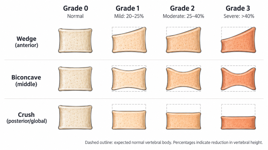
Figure 1. Genant semiquantitative grading of vertebral compression deformities according to the reduction in vertebral height: grade 0, normal; grade 1 (mild), 20–25%; grade 2 (moderate), 25–40%; grade 3 (severe), >40%. Wedge (anterior), biconcave (middle) and crush (posterior or global) configurations are shown.
Treatment
Traumatic A1 fractures are treated with early mobilization and analgesia; a brace may be used for comfort, and standing radiographs at 6–12 weeks confirm stability. For osteoporotic fractures, nonoperative care is first-line: multimodal analgesia with minimal opioid use, early mobilization, physiotherapy and fall prevention. Prolonged bed rest should be avoided. Starting or optimizing anti-osteoporotic treatment is essential, and anabolic agents are increasingly favored in patients at very high fracture risk. Referral to a fracture liaison service improves long-term treatment adherence.
Vertebral augmentation remains controversial. Sham-controlled trials have not shown a consistent benefit of vertebroplasty over a sham procedure [3], and current guidance does not support routine use [4]. Augmentation may be considered in selected patients with acute fractures, MRI edema and severe pain despite optimal medical care, particularly hospitalized patients. OF4 and OF5 fractures, neurological deficits and progressive collapse require stabilization, often with cement-augmented pedicle screws.