Figures
Overview
High-energy trauma, such as falls from height and traffic accidents, predominates in young adults. Low-energy falls cause most fractures in older adults with osteoporosis or ankylosed spines. Associated injuries are common: calcaneal and lower-limb fractures after falls, intra-abdominal injuries after seatbelt mechanisms, and noncontiguous spinal fractures.
Clinical Presentation
Patients present with axial pain, focal tenderness, a palpable step-off or interspinous gap and, after seatbelt injuries, abdominal wall ecchymosis. A complete neurological examination according to the International Standards (ISNCSCI) is mandatory. It must include perianal sensation, voluntary anal contraction and the bulbocavernosus reflex to detect conus medullaris or cauda equina injury. In polytrauma, the spine is assessed within the ATLS framework, and spinal motion restriction is maintained until injury is excluded.
Imaging
Multidetector CT of the whole spine is the standard in high-energy trauma. It defines fracture morphology, canal compromise and posterior element injury. MRI is indicated for neurological deficits, for suspected posterior ligamentous complex (PLC) injury when CT is equivocal, and for distinguishing acute from chronic fractures. Upright radiographs reveal kyphosis progression under load in neurologically intact patients considered for nonoperative treatment.
CT signs of PLC injury include widening of the interspinous distance, facet diastasis or perched facets, and avulsion of the spinous process tips. On MRI, STIR hyperintensity of the interspinous ligament is sensitive but not specific; discontinuity of the supraspinous ligament or ligamentum flavum is a more reliable sign of disruption.
Classification
The AO Spine thoracolumbar classification describes morphology (type A, compression; type B, tension-band failure; type C, translation), neurological status (N0–N4, NX) and case-specific modifiers (M1, indeterminate PLC injury; M2, patient-specific comorbidity such as ankylosing spondylitis) (Figure 1) [1]. The Thoracolumbar AO Spine Injury Score (TL AOSIS) converts these elements into points: scores ≤3 favor nonoperative treatment, scores >5 favor surgery, and scores of 4–5 are equivocal [2]. The older TLICS uses morphology, PLC integrity and neurology in a similar way.
Neurological grading is practical: N0, intact; N1, transient deficit; N2, radicular symptoms; N3, incomplete cord or cauda equina injury; N4, complete cord injury; NX, patient cannot be examined. Minor process fractures (A0) do not affect stability and need symptomatic care only.
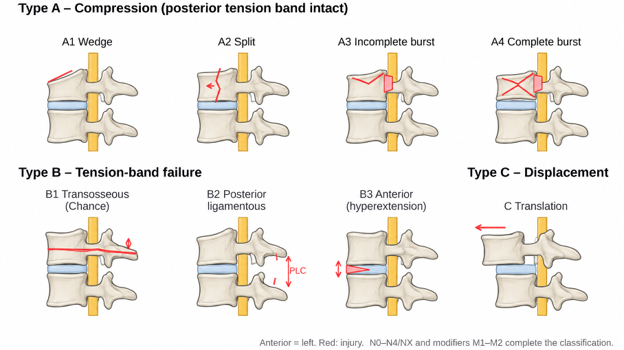
Figure 1. Schematic overview of the AO Spine thoracolumbar injury classification. Type A injuries are compression injuries of the vertebral body with an intact posterior tension band (A1 wedge, A2 split, A3 incomplete burst, A4 complete burst). Type B injuries involve failure of the posterior (B1, B2) or anterior (B3) tension band. Type C injuries show displacement or translation in any plane.
Treatment
Most type A injuries without neurological deficit and with an intact PLC are treated nonoperatively with early mobilization. Bracing is optional; a randomized equivalence trial showed no advantage of an orthosis over mobilization alone in stable burst fractures [4]. Adequate analgesia, thromboprophylaxis and serial upright radiographs are essential.
Surgery aims to decompress neural elements, restore stability and alignment, and allow early mobilization [3]. Posterior pedicle screw fixation is the workhorse. Short-segment constructs with screws in the fractured vertebra are adequate for most injuries; long-segment or combined anterior reconstruction is reserved for severe comminution or poor bone quality. Percutaneous fixation reduces soft-tissue damage in neurologically intact patients. In the low lumbar spine, maintaining lordosis and avoiding fusion to the sacrum are key goals.
When short-segment percutaneous fixation is used for type A injuries, fusion can often be omitted, and implant removal after healing may restore segmental motion; its routine use remains debated. Decompression is added when neural compression persists after reduction.
Surgical Indications
• Neurological deficit with ongoing neural compression
• Type B and C injuries (tension-band disruption or translation)
• TL AOSIS >5 (or TLICS ≥5)
• Progressive neurological deficit or deformity during nonoperative care
• Polytrauma or patient factors precluding mobilization
Prognosis
Neurologically intact patients generally recover well with either treatment, and residual kyphosis correlates poorly with pain and function. Neurological recovery depends mainly on the initial deficit; cauda equina injuries have a better prognosis than conus or cord injuries.
Differential Diagnosis
• Osteoporotic insufficiency fractures
• Pathological fractures (metastasis, myeloma, infection)
• Developmental wedging (e.g., Scheuermann disease) and limbus vertebra
References
1. Vaccaro AR, Oner C, Kepler CK, et al. AOSpine thoracolumbar spine injury classification system: fracture description, neurological status, and key modifiers. Spine (Phila Pa 1976). 2013;38(23):2028-37. doi:10.1097/BRS.0b013e3182a8a381
2. Kepler CK, Vaccaro AR, Schroeder GD, et al. The Thoracolumbar AOSpine Injury Score. Global Spine J. 2016;6(4):329-34.
3. Vaccaro AR, Schroeder GD, Kepler CK, et al. The surgical algorithm for the AOSpine thoracolumbar spine injury classification system. Eur Spine J. 2016;25(4):1087-94.
4. Bailey CS, Urquhart JC, Dvorak MF, et al. Orthosis versus no orthosis for the treatment of thoracolumbar burst fractures without neurologic injury: a multicenter prospective randomized equivalence trial. Spine J. 2014;14(11):2557-64.