Figures
Overview
Isthmic spondylolisthesis is the forward slip of a vertebra secondary to a defect of the pars interarticularis (Wiltse type II) [3]. The defect, bilateral in most cases, usually develops in childhood as a fatigue fracture caused by repetitive extension loading and is present in about 6% of adults [4]; most cases involve L5–S1. Slip progression in adults is uncommon. Symptoms usually appear in the third to fifth decades, when disc degeneration at the slipped level produces mechanical back pain and foraminal stenosis. A high pelvic incidence is associated with greater slips.
Clinical Presentation
Patients present with mechanical low back pain, radicular pain or both. Radicular symptoms usually involve the L5 root, which is compressed in the L5–S1 foramen by the fibrocartilaginous mass at the pars defect, the bulging disc and the loss of foraminal height. Neurogenic claudication occurs with higher-grade slips. A step-off may be palpable; neurological deficits are usually mild. Hamstring tightness and the typical crouched gait are more characteristic of adolescents with high-grade slips than of adults.
Imaging
Standing lateral radiographs establish the slip grade and slip angle, and flexion-extension views assess segmental mobility (Figure 1). Meyerding grades are I (0–25%), II (26–50%), III (51–75%), IV (76–100%) and V (>100%, spondyloptosis); grades I–II are considered low grade. CT best demonstrates the pars defects; oblique radiographs, which show the classic "Scottie dog collar", have largely been replaced by CT. MRI evaluates foraminal stenosis, root compression and disc degeneration, including at adjacent levels. Full-length standing radiographs with spinopelvic parameters are recommended for high-grade slips and surgical planning.
In high-grade slips, the slip angle (lumbosacral kyphosis) and sacropelvic balance matter more than the slip percentage. The Spinal Deformity Study Group classification separates low-grade slips by pelvic incidence and high-grade slips by sacropelvic and global spinal balance; unbalanced high-grade slips are the strongest argument for partial reduction.
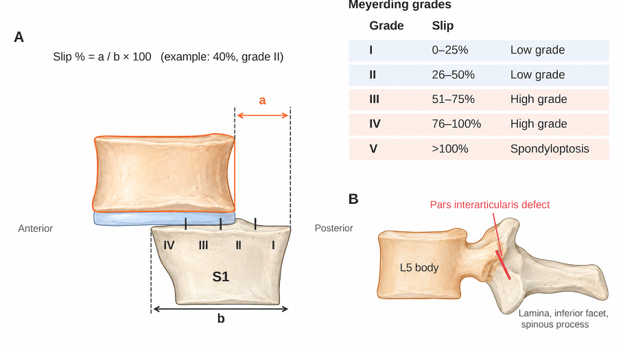
Figure 1. (A) Meyerding grading of spondylolisthesis: the percentage of anterior translation of L5 relative to the superior endplate of S1 (a/b × 100) determines grades I–V. (B) Lateral schematic of the pars interarticularis defect separating the vertebral body, pedicles and superior facets from the lamina, inferior facets and spinous process.
Treatment
Nonoperative management is first-line for most patients: activity modification, analgesics, physiotherapy focusing on core stabilization and flexion-based exercises, and transforaminal epidural steroid injections for radicular pain [1]. Most patients with low-grade slips can be managed without surgery.
Surgery is considered after failure of at least 3–6 months of structured nonoperative care. In a randomized trial, posterolateral fusion gave better pain and functional outcomes than an exercise program [2]. Decompression alone (Gill procedure) is generally avoided in adults because it increases instability. Instrumented fusion is the standard and usually includes L5–S1 only; extension to L4 is considered when the L4–L5 disc is degenerated or when a high-grade slip requires additional fixation.
Interbody techniques (TLIF, PLIF or ALIF) restore disc and foraminal height, provide indirect decompression and improve fusion rates [5]. Minimally invasive TLIF achieves similar results with less blood loss in experienced hands. Direct foraminal decompression is added when root compression persists. Direct pars repair is reserved for young adults with symptomatic spondylolysis, minimal slip and a healthy disc. In high-grade slips, partial reduction improves sagittal alignment and fusion biology, but aggressive reduction increases the risk of L5 root injury.
Surgical Indications
• Persistent disabling pain or radiculopathy despite adequate nonoperative care
• Progressive neurological deficit or cauda equina syndrome
• Documented slip progression or symptomatic high-grade slip
• Segmental instability with concordant symptoms
Prognosis
Most patients improve substantially after fusion, with high union rates when interbody fusion is used [5]. L5 root deficits after reduction of high-grade slips are usually transient. Patients with predominantly radicular symptoms tend to have better outcomes than those with axial pain alone. Smoking, high-grade slips and poor bone quality increase the risk of pseudarthrosis. Adjacent segment degeneration may develop in the long term.
Differential Diagnosis
• Degenerative spondylolisthesis (usually L4–L5, intact pars, facet arthropathy, older patients)
• Dysplastic spondylolisthesis (Wiltse type I)
• Acute traumatic pars fracture
• Disc herniation, facet cysts or foraminal stenosis from other causes
References
1. Kreiner DS, Baisden J, Mazanec DJ, et al. Guideline summary review: an evidence-based clinical guideline for the diagnosis and treatment of adult isthmic spondylolisthesis. Spine J. 2016;16(12):1478-85.
2. Möller H, Hedlund R. Surgery versus conservative management in adult isthmic spondylolisthesis—a prospective randomized study: part 1. Spine (Phila Pa 1976). 2000;25(13):1711-5.
3. Wiltse LL, Newman PH, Macnab I. Classification of spondylolisis and spondylolisthesis. Clin Orthop Relat Res. 1976;(117):23-9.
4. Fredrickson BE, Baker D, McHolick WJ, Yuan HA, Lubicky JP. The natural history of spondylolysis and spondylolisthesis. J Bone Joint Surg Am. 1984;66(5):699-707.
5. Noorian S, Sorensen K, Cho W. A systematic review of clinical outcomes in surgical treatment of adult isthmic spondylolisthesis. Spine J. 2018;18(8):1441-54.