Figures
Overview
Adult spinal deformity (ASD) includes de novo degenerative scoliosis, progression of adolescent idiopathic scoliosis, iatrogenic deformity such as flatback, and primary sagittal malalignment [4]. Radiographic scoliosis is present in a large proportion of elderly people, and symptomatic ASD causes disability comparable to that of major chronic diseases. Sagittal alignment, rather than coronal curve magnitude, correlates most strongly with pain and disability [2].
Clinical Presentation
Patients present with axial back pain, radiculopathy or neurogenic claudication, often from foraminal stenosis in the concavity of the lumbosacral fractional curve, and progressive deformity. Sagittal malalignment causes forward trunk inclination, fatigue and difficulty standing or walking. Compensation through thoracic hypokyphosis, pelvic retroversion, hip extension and knee flexion can mask the true deformity. Frailty, osteoporosis, nutrition and comorbidities must be assessed because they strongly influence surgical risk.
Imaging
Full-length standing posteroanterior and lateral radiographs including the femoral heads, or biplanar low-dose imaging, are essential. Key parameters are (Figure 1):
• Sagittal vertical axis (SVA): horizontal offset of the C7 plumb line from the posterosuperior corner of S1
• Pelvic incidence (PI), a fixed morphological parameter, equal to pelvic tilt (PT) plus sacral slope (SS)
• Lumbar lordosis (LL) and the PI–LL mismatch
• T1 pelvic angle and coronal balance (C7 plumb line to the central sacral vertical line)
Supine or bending films assess flexibility. MRI evaluates neural compression. CT assists planning and, with densitometry, the assessment of bone quality; low vertebral Hounsfield units indicate a high risk of screw loosening and junctional failure.
Classification
The SRS–Schwab classification combines the coronal curve type (thoracic, lumbar, double or none) with three sagittal modifiers: PI–LL (<10°, 10–20°, >20°), PT (<20°, 20–30°, >30°) and SVA (<4 cm, 4–9.5 cm, >9.5 cm) [1]. Higher modifier grades correlate with worse health-related quality of life, measured with instruments such as the Oswestry Disability Index and SRS-22r. Alignment targets have evolved from fixed thresholds (PI–LL <10°, PT <20°, SVA <5 cm) to age-adjusted and proportional targets. The Global Alignment and Proportion (GAP) score, based on pelvic incidence, predicts mechanical complications after surgery [3].
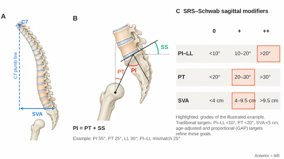
Figure 1. Sagittal alignment in adult spinal deformity. (A) Whole-spine alignment with the C7 plumb line and sagittal vertical axis (SVA). (B) Spinopelvic parameters: pelvic incidence (PI), pelvic tilt (PT) and sacral slope (SS), where PI = PT + SS. (C) SRS–Schwab sagittal modifiers; the highlighted cells correspond to the illustrated example. LL, lumbar lordosis.
Treatment
Nonoperative care, including physiotherapy, analgesics, injections, bone-health optimization and weight control, is appropriate for patients with mild symptoms or high surgical risk. Durable improvement is uncommon in patients with marked malalignment.
Surgical planning starts with the patient's goals and risk profile. Frailty indices, sarcopenia, nutrition and smoking status help stratify risk. The strategy depends on where the symptoms arise: isolated radicular symptoms with a stable, flexible curve may respond to decompression and short fusion, whereas global sagittal malalignment requires long fusion with realignment. In elderly patients, age-adjusted targets avoid overcorrection, which increases the risk of proximal junctional failure.
Correction techniques include posterior column osteotomies (Smith-Petersen or Ponte), pedicle subtraction osteotomy (about 30° of lordosis per level), vertebral column resection for rigid multiplanar deformities, and anterior or lateral interbody fusion to restore segmental lordosis. Long constructs to the pelvis are anchored with iliac or S2-alar-iliac screws. Proximal junctional kyphosis is reduced by careful selection of the upper instrumented vertebra, avoiding overcorrection, preserving posterior ligaments, tethers and cement augmentation.
Preoperative optimization of bone quality, for example with anabolic agents, is increasingly used. Staging long procedures and multidisciplinary perioperative care reduce complications in elderly patients.
Surgical Indications
• Persistent pain and disability despite adequate nonoperative care
• Progressive coronal or sagittal deformity
• Neurogenic claudication or radiculopathy with deformity-related stenosis
• Severe sagittal malalignment limiting standing or walking
• Medical fitness, adequate bone quality and realistic expectations after shared decision-making
Prognosis
In selected patients, surgery provides greater improvement in pain and function than nonoperative care [5]. Complication rates remain high, including proximal junctional kyphosis (a proximal junctional angle of ≥10° that is ≥10° greater than before surgery), rod fracture, pseudarthrosis, infection and neurological deficit, and revision surgery is common. Restoring alignment proportional to the patient's pelvic morphology reduces mechanical failure [3].
Differential Diagnosis
• Osteoporotic vertebral fractures with kyphosis
• Camptocormia of neuromuscular origin (Parkinson disease, myopathies)
• Hip flexion contracture or hip osteoarthritis
• Ankylosing spondylitis and iatrogenic flatback
References
1. Schwab F, Ungar B, Blondel B, et al. Scoliosis Research Society-Schwab adult spinal deformity classification: a validation study. Spine (Phila Pa 1976). 2012;37(12):1077-82.
2. Glassman SD, Bridwell K, Dimar JR, Horton W, Berven S, Schwab F. The impact of positive sagittal balance in adult spinal deformity. Spine (Phila Pa 1976). 2005;30(18):2024-9.
3. Yilgor C, Sogunmez N, Boissiere L, et al. Global Alignment and Proportion (GAP) score: development and validation of a new method of analyzing spinopelvic alignment to predict mechanical complications after adult spinal deformity surgery. J Bone Joint Surg Am. 2017;99(19):1661-72. doi:10.2106/JBJS.16.01594
4. Diebo BG, Shah NV, Boachie-Adjei O, et al. Adult spinal deformity. Lancet. 2019;394(10193):160-72. doi:10.1016/S0140-6736(19)31125-0
5. Kelly MP, Lurie JD, Yanik EL, et al. Operative versus nonoperative treatment for adult symptomatic lumbar scoliosis. J Bone Joint Surg Am. 2019;101(4):338-52.