Figures
Overview
Congenital scoliosis affects roughly 0.5–1 per 1000 live births and results from vertebral malformations arising during somite formation and segmentation in the first 4–6 weeks of gestation. Anomalies are classified as failures of formation (hemivertebrae, wedge vertebrae), failures of segmentation (unilateral bars, block vertebrae) or mixed forms (Figure 1). Because the same embryological window affects other organ systems, associated anomalies are common: intraspinal anomalies (tethered cord, diastematomyelia, syringomyelia) occur in about one-third of patients and cardiac or genitourinary anomalies in about one-fifth to one-quarter [2]. Syndromic associations include VACTERL, Klippel-Feil, Goldenhar and Alagille syndromes and spondylocostal dysostosis.
Natural History
Progression depends on the type and location of the anomaly and on remaining growth [1]. A unilateral unsegmented bar with a contralateral hemivertebra has the worst prognosis, followed by an isolated unilateral bar and double fully segmented hemivertebrae. Block vertebrae and incarcerated hemivertebrae rarely progress. Congenital kyphosis caused by anterior failure of formation carries a risk of paraplegia and requires early surgical treatment. Progression is fastest during periods of rapid growth, in the first years of life and during the adolescent growth spurt; thoracolumbar and lumbosacral anomalies produce the most visible deformities.
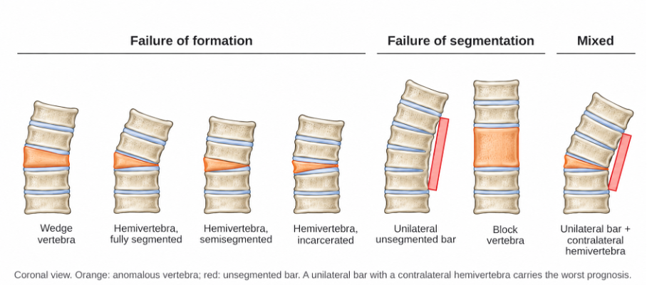
Figure 1. Classification of congenital scoliosis. Failure of formation: wedge vertebra and hemivertebra (fully segmented, semisegmented, incarcerated). Failure of segmentation: unilateral unsegmented bar and block vertebra. Mixed: unilateral bar with a contralateral hemivertebra, which carries the worst prognosis.
Clinical Presentation
Most cases are sporadic, and a family history is usually absent. Many are detected on prenatal ultrasound or incidental radiographs. Children present with trunk asymmetry, shoulder or pelvic obliquity, or a short trunk. Cutaneous stigmata over the spine (hair patch, dimple, hemangioma), foot deformities, leg-length discrepancy and bladder dysfunction suggest an intraspinal anomaly. A full neurological examination and cardiac and renal assessment are required [3].
Imaging
Standing radiographs are used for diagnosis and follow-up, measuring the Cobb angle between the same end vertebrae on serial images, because measurement variability is high in young children with complex anomalies. Low-dose CT with three-dimensional reconstruction defines the anatomy for surgical planning. Whole-spine MRI is mandatory before surgery and whenever neurological signs are present [6]. Renal ultrasound and echocardiography screen for associated anomalies. In children with fused ribs or a short thorax, CT, pulmonary function testing where feasible and assessment of thoracic growth define thoracic insufficiency syndrome.
Treatment
Curves with a low risk of progression are observed with radiographs every 4–6 months during rapid growth. Bracing does not control the congenital curve itself, although it may be used for compensatory curves.
Surgery aims to prevent progression and achieve a balanced spine with minimal loss of growth [5]. Early posterior hemivertebra resection with short-segment instrumentation, ideally before secondary structural changes develop in early childhood, provides good correction for isolated hemivertebrae [4]; excision before about 5–6 years of age allows short fusions and prevents compensatory structural curves.
In situ fusion or convex hemiepiphysiodesis is an option for selected curves and is most effective before the curve becomes large and rigid. Growth-friendly techniques, such as traditional or magnetically controlled growing rods and rib-based distraction, are used for long curves or thoracic insufficiency syndrome with fused ribs. Severe rigid curves in older children may require vertebral column resection. Symptomatic intraspinal anomalies are addressed before or at the time of correction. Neuromonitoring is essential, because neurological risk is higher than in idiopathic scoliosis.
Surgical Indications
• Documented progression
• Anomalies with a poor natural history (unilateral bar with or without a contralateral hemivertebra)
• Lumbosacral hemivertebra causing trunk shift or pelvic obliquity
• Thoracic insufficiency syndrome
• Neurological deficit related to the deformity
Prognosis
Early, appropriately selected surgery yields good long-term results. Complications include neurological injury, implant failure, junctional deformity, crankshaft progression after posterior-only fusion in very young children and, after long early fusions, reduced trunk height and pulmonary function. Untreated progressive curves can lead to severe deformity and cardiopulmonary compromise. Follow-up should continue until skeletal maturity, with attention to junctional segments.
Differential Diagnosis
• Early-onset idiopathic scoliosis
• Neuromuscular scoliosis
• Syndromic scoliosis (e.g., neurofibromatosis, Marfan syndrome)
• Skeletal dysplasias
References
1. McMaster MJ, Ohtsuka K. The natural history of congenital scoliosis. A study of two hundred and fifty-one patients. J Bone Joint Surg Am. 1982;64(8):1128-47.
2. Basu PS, Elsebaie H, Noordeen MH. Congenital spinal deformity: a comprehensive assessment at presentation. Spine (Phila Pa 1976). 2002;27(20):2255-9.
3. Hedequist D, Emans J. Congenital scoliosis. J Am Acad Orthop Surg. 2004;12(4):266-75.
4. Yaszay B, O'Brien M, Shufflebarger HL, et al. Efficacy of hemivertebra resection for congenital scoliosis: a multicenter retrospective comparison of three surgical techniques. Spine (Phila Pa 1976). 2011;36(24):2052-60.
5. Pahys JM, Guille JT. What's new in congenital scoliosis? J Pediatr Orthop. 2018;38(3):e172-9.
6. Mackel CE, Jada A, Samdani AF, et al. A comprehensive review of the diagnosis and management of congenital scoliosis. Childs Nerv Syst. 2018;34(11):2155-71.