Figures
Overview
In ankylosing spondylitis (AS) and diffuse idiopathic skeletal hyperostosis (DISH), the risk of spinal fracture is several times higher than in the general population. Long fused segments convert the spine into a rigid, brittle lever. Fractures occur after low-energy trauma, such as a fall from standing height, and typically extend through all three columns; they are highly unstable, often AO Spine type B3 or C injuries [1,3]. In AS, fractures often pass through the ossified disc space and most commonly affect the lower cervical spine and cervicothoracic junction; osteoporosis is frequent. In DISH, fractures more often traverse the vertebral body. Patients are often elderly with multiple comorbidities, and morbidity and mortality are substantially higher than in non-ankylosed spines [2].
Clinical Presentation
Any new pain after even minor trauma in an ankylosed spine is a fracture until proven otherwise. Patients may report a change in posture or "new mobility". Neurological deficits are frequent and may appear or worsen after a delay because of displacement or epidural hematoma. Patients must be immobilized in their pre-injury posture, using pillows if needed. Forcing a kyphotic neck into a standard collar or a flat supine position can displace the fracture and cause neurological deterioration. Elderly patients with DISH often sustain cervical hyperextension injuries after falls onto the face. Fixed cervical kyphosis and reduced chest expansion make airway management difficult; awake fiberoptic intubation is often required.
Imaging
Radiographs miss many fractures because of osteopenia and ossification. Minimally displaced hyperextension fractures may also appear reduced on supine imaging, giving a false impression of stability. CT of the entire spine is mandatory, as noncontiguous fractures are common. MRI with STIR sequences detects occult fractures, disc-space injuries and epidural hematoma and should be obtained when neurological symptoms are present or pain persists despite negative CT.
Distinguishing features (Figure 1) [5]:
• AS: marginal syndesmophytes ("bamboo spine"), facet and sacroiliac joint ankylosis, osteoporosis, younger age at onset
• DISH: flowing ossification along the anterolateral aspect of at least four contiguous vertebrae, preserved disc height, no sacroiliac ankylosis, older age and metabolic comorbidity
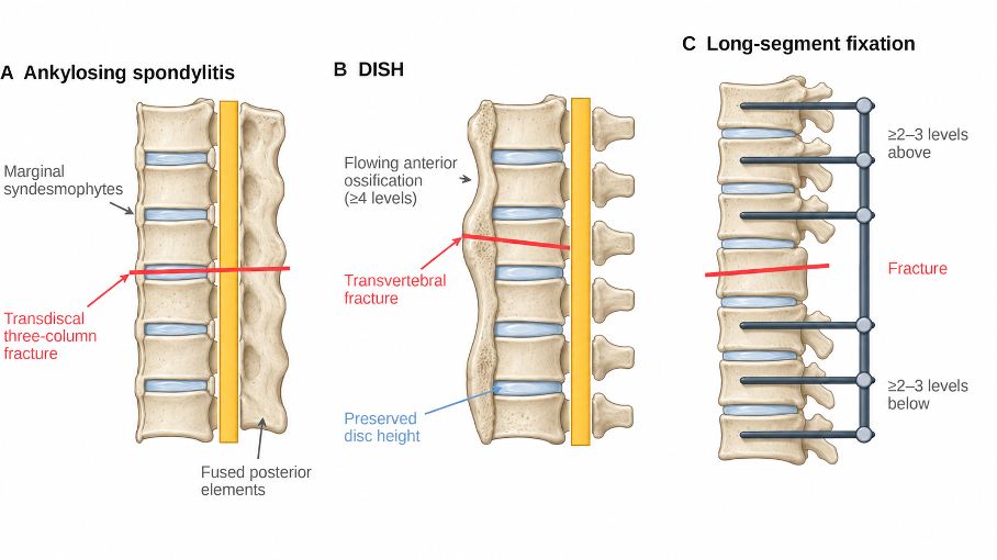
Figure 1. Fractures in ankylosing spinal disorders. (A) Ankylosing spondylitis: marginal syndesmophytes, fused posterior elements and a transdiscal fracture extending through all three columns. (B) Diffuse idiopathic skeletal hyperostosis (DISH): flowing anterior ossification with preserved disc height and a transvertebral fracture. (C) Principle of long-segment posterior fixation spanning at least two to three levels above and below the fracture.
Treatment
Most fractures require surgical stabilization [3,4]. Posterior long-segment instrumentation extending at least two to three levels above and below the fracture is the standard. Percutaneous techniques reduce blood loss and soft-tissue morbidity. In osteoporotic bone, cement augmentation, additional fixation points or laminar hooks may improve construct stability. The goal is to restore pre-injury alignment rather than to correct the chronic deformity. Anterior-only constructs are prone to failure; combined anterior–posterior surgery is reserved for selected cervical fractures with gross displacement or anterior compression. Nonoperative treatment with a halo or brace is reserved for selected nondisplaced fractures in patients unfit for surgery and carries a high risk of displacement and complications.
Positioning is critical: the pre-injury kyphosis must be supported with bolsters or a tilted frame, and alignment is checked with fluoroscopy before incision. Constructs should not end at the fracture-adjacent junction, and longer fixation is preferred in osteoporotic bone. Early surgery allows mobilization and reduces complications related to immobility.
Surgical Indications
• Virtually all unstable fractures, with or without neurological deficit
• Neurological deficit or deterioration
• Epidural hematoma with neurological compromise (emergency decompression)
• Failure of nonoperative treatment (displacement, pseudarthrosis)
Prognosis
Union rates after long-segment fixation are high, but perioperative complications such as pneumonia, delirium, thromboembolism and wound problems are common in these often elderly and frail patients. Delayed diagnosis and secondary neurological deterioration are the main avoidable causes of poor outcome. Mortality is highest in elderly patients with cervical fractures and neurological deficits. Postoperative care should include early mobilization, pulmonary physiotherapy and treatment of osteoporosis.
Differential Diagnosis
• Andersson lesion (spondylodiscitis or pseudarthrosis in AS)
• Infectious spondylodiscitis
• Osteoporotic or pathological fractures
• Progressive kyphotic deformity without acute fracture
References
1. Westerveld LA, Verlaan JJ, Oner FC. Spinal fractures in patients with ankylosing spinal disorders: a systematic review of the literature on treatment, neurological status and complications. Eur Spine J. 2009;18(2):145-56.
2. Caron T, Bransford R, Nguyen Q, et al. Spine fractures in patients with ankylosing spinal disorders. Spine (Phila Pa 1976). 2010;35(11):E458-64.
3. Reinhold M, Knop C, Kneitz C, Disch A. Spine fractures in ankylosing diseases: recommendations of the Spine Section of the German Society for Orthopaedics and Trauma (DGOU). Global Spine J. 2018;8(2 Suppl):56S-68S.
4. Rustagi T, Drazin D, Oner C, et al. Fractures in spinal ankylosing disorders: a narrative review of disease and injury types, treatment techniques, and outcomes. J Orthop Trauma. 2017;31 Suppl 4:S57-74.
5. Resnick D, Niwayama G. Radiographic and pathologic features of spinal involvement in diffuse idiopathic skeletal hyperostosis (DISH). Radiology. 1976;119(3):559-68.