Figures
Overview
Primary malignant tumors can be cured by resection that follows oncological principles, whereas metastatic disease is treated to relieve pain, preserve neurological function and maintain stability. Tissue diagnosis and multidisciplinary planning precede definitive treatment.
Primary Spine Tumors
Clinical Presentation
Fewer than 10% of primary bone tumors arise in the spine. Benign lesions, such as osteoid osteoma, osteoblastoma and aneurysmal bone cyst, are more common in young patients and often involve the posterior elements. Malignant tumors, such as chordoma, chondrosarcoma, osteosarcoma and Ewing sarcoma, more often involve the vertebral body. Multiple myeloma and solitary plasmacytoma are the most common primary malignancies of the spine.
The most common symptom is back pain that increases at night and is not relieved by rest. Osteoid osteoma may present with painful scoliosis. Radiculopathy or myelopathy may accompany pain. Slow-growing tumors such as chordoma and chondrosarcoma are often diagnosed late.
Imaging and Biopsy
Radiographs may be normal early, because lytic lesions become visible only after substantial bone loss. CT shows bone destruction and matrix mineralization, and MRI shows soft-tissue extension and neural involvement.
Definitive diagnosis requires biopsy, which should be planned with the treating surgeon. The biopsy tract must lie within the planned resection so that it can be excised en bloc; an inappropriate biopsy contaminates tissue planes and increases the risk of local recurrence [1].
Staging and Treatment
Enneking staging defines the surgical margin required. The Weinstein–Boriani–Biagini (WBB) system maps tumor extent in 12 radiating zones and five layers and guides the choice of resection (Figure 1A) [2]. For primary malignant and locally aggressive benign (Enneking stage 3) tumors, the aim is en bloc resection with appropriate margins, which reduces local recurrence and mortality [3]. Benign latent or active lesions are treated with intralesional excision or percutaneous ablation.
When en bloc resection is not feasible, the usual alternative is intralesional excision followed by radiotherapy; high-dose proton or carbon-ion therapy is often used for chordoma and chondrosarcoma. Osteosarcoma and Ewing sarcoma require chemotherapy as part of multimodal treatment, whereas myeloma, plasmacytoma and lymphoma are radiosensitive and are treated mainly with radiotherapy and systemic therapy. Stability is restored with instrumented fusion and anterior column reconstruction.
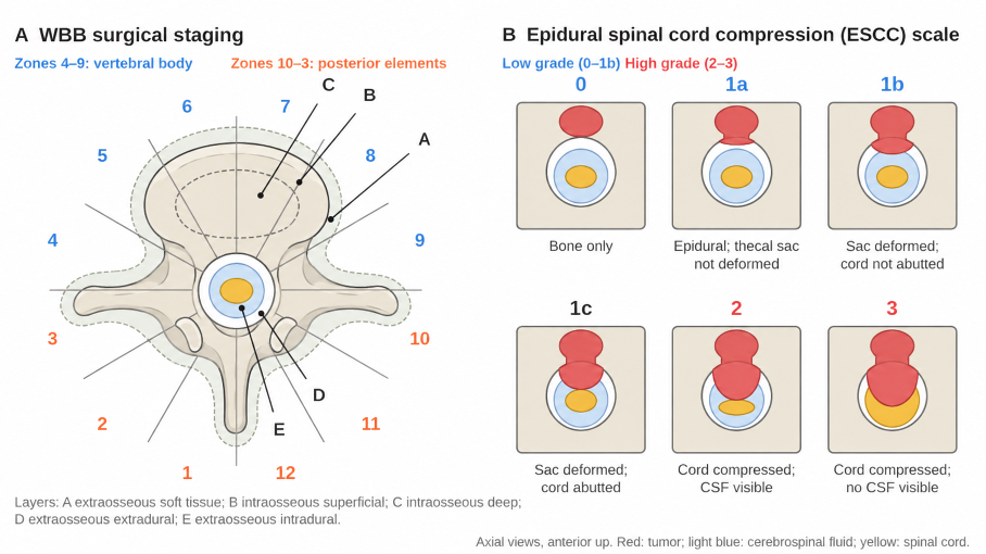
Figure 1. (A) Weinstein–Boriani–Biagini (WBB) surgical staging. In the axial plane, the vertebra is divided into 12 radiating zones, numbered clockwise, and five layers: A, extraosseous soft tissue; B, intraosseous superficial; C, intraosseous deep; D, extraosseous extradural; E, extraosseous intradural. Zones 4–9 correspond to the vertebral body and zones 10–3 to the posterior elements. (B) Spine Oncology Study Group epidural spinal cord compression (ESCC) scale. Grades 0–1b are low grade, grade 1c (cord abutment without compression) is intermediate, and grades 2–3 are high grade.
Metastatic Spine Tumors
Clinical Presentation
The spine is the most common site of skeletal metastasis; autopsy series show spinal metastases in 30–70% of patients with cancer. Lung, breast, prostate, kidney, thyroid and colorectal cancers are the most frequent sources. About 80% of lesions involve the vertebral body, and about 70% occur in the thoracic spine.
Pain is the most common symptom. Biological pain is worse at night and at rest, whereas mechanical pain with movement or loading suggests instability. Metastatic epidural spinal cord compression (MESCC) develops in 5–10% of patients with cancer and can cause neurological deficit.
Diagnostic Evaluation
Whole-spine MRI is the gold standard for detecting epidural compression, which is graded with the Spine Oncology Study Group ESCC scale (Figure 1B) [4]. The Spinal Instability Neoplastic Score (SINS) assesses mechanical stability: 0–6 stable, 7–12 potentially unstable and 13–18 unstable; a score of 7 or more warrants surgical consultation [5]. PET-CT is valuable for systemic staging, and biopsy is needed when the primary tumor is unknown.
Treatment
Decisions follow the NOMS framework, which integrates neurological, oncological (radiosensitivity), mechanical and systemic considerations [6]. Radiotherapy and systemic therapy (chemotherapy, immunotherapy, hormonal or targeted agents) are the basis of treatment. In a randomized trial, stereotactic body radiotherapy (SBRT; 24 Gy in 2 fractions) produced a higher complete pain response than conventional radiotherapy (20 Gy in 5 fractions) [7]. SBRT also achieves durable local control in tumors resistant to conventional radiotherapy [6,8].
The standard operation is decompression and stabilization. In a randomized trial of selected patients with MESCC, decompressive surgery followed by radiotherapy enabled 84% of patients to walk after treatment, compared with 57% after radiotherapy alone [9]. Separation surgery decompresses the cord to allow safe postoperative SBRT, and en bloc resection is reserved for selected solitary metastases [8]. Vertebroplasty, kyphoplasty and percutaneous fixation relieve pain in patients with limited life expectancy.
Surgical Indications
• Intractable pain despite nonoperative treatment
• Mechanical instability (SINS 13 or higher; consider surgery for 7–12)
• Neurological deficit or high-grade MESCC, particularly in radioresistant tumors
Prognosis
In metastatic disease, the aim is to improve quality of life rather than to cure. Survival depends on tumor histology, the burden of systemic disease and performance status. Scoring systems such as the revised Tokuhashi score help match treatment intensity to expected survival [10]; surgery is generally considered when survival is expected to exceed 3 months.
Complications
• Neurological deficit and massive bleeding; preoperative embolization reduces blood loss in hypervascular tumors such as renal and thyroid metastases
• Wound infection and dehiscence, particularly after radiotherapy
• Implant failure and mechanical instability
• Cement leakage after augmentation
• Systemic complications such as pulmonary embolism and pneumonia
• Local recurrence after inadequate margins in primary tumors
Multidisciplinary follow-up by oncology, radiation oncology, orthopedic and neurosurgical teams is essential.
Differential Diagnosis
• Spondylodiscitis (disc involvement; tumors usually spare the disc)
• Osteoporotic compression fracture
• Multiple myeloma, plasmacytoma and lymphoma
• Hemangioma, bone island and Paget disease
References
1. Charest-Morin R, Fisher CG, Sahgal A, et al. Primary bone tumor of the spine—an evolving field: what a general spine surgeon should know. Global Spine J. 2019;9(1 Suppl):108S-116S.
2. Boriani S, Weinstein JN, Biagini R. Primary bone tumors of the spine: terminology and surgical staging. Spine (Phila Pa 1976). 1997;22(9):1036-44.
3. Fisher CG, Saravanja DD, Dvorak MF, et al. Surgical management of primary bone tumors of the spine: validation of an approach to enhance cure and reduce local recurrence. Spine (Phila Pa 1976). 2011;36(10):830-6.
4. Bilsky MH, Laufer I, Fourney DR, et al. Reliability analysis of the epidural spinal cord compression scale. J Neurosurg Spine. 2010;13(3):324-8.
5. Fisher CG, DiPaola CP, Ryken TC, et al. A novel classification system for spinal instability in neoplastic disease: an evidence-based approach and expert consensus from the Spine Oncology Study Group. Spine (Phila Pa 1976). 2010;35(22):E1221-9. doi:10.1097/BRS.0b013e3181e16ae2
6. Laufer I, Rubin DG, Lis E, et al. The NOMS framework: approach to the treatment of spinal metastatic tumors. Oncologist. 2013;18(6):744-51. doi:10.1634/theoncologist.2012-0293
7. Sahgal A, Myrehaug SD, Siva S, et al. Stereotactic body radiotherapy versus conventional external beam radiotherapy in patients with painful spinal metastases: an open-label, multicentre, randomised, controlled, phase 2/3 trial. Lancet Oncol. 2021;22(7):1023-33. doi:10.1016/S1470-2045(21)00196-0
8. Barzilai O, Fisher CG, Bilsky MH. State of the art treatment of spinal metastatic disease. Neurosurgery. 2018;82(6):757-69.
9. Patchell RA, Tibbs PA, Regine WF, et al. Direct decompressive surgical resection in the treatment of spinal cord compression caused by metastatic cancer: a randomised trial. Lancet. 2005;366(9486):643-8. doi:10.1016/S0140-6736(05)66954-1
10. Tokuhashi Y, Matsuzaki H, Oda H, Oshima M, Ryu J. A revised scoring system for preoperative evaluation of metastatic spine tumor prognosis. Spine (Phila Pa 1976). 2005;30(19):2186-91.