Figures
Overview
The posterior and middle columns fail in tension, and the anterior column fails in compression or, less often, in tension. In the AO Spine system they are type B1 (transosseous disruption of the posterior tension band, the classic "bony Chance" fracture) or type B2 (ligamentous or osseoligamentous disruption) [1,2]. Most occur at T12–L2 in adults and in the mid-lumbar spine in children restrained with lap belts, in whom the belt rides above the pelvis and the flexion axis lies against the abdominal wall.
Clinical Presentation
Patients present with back pain and a palpable gap between spinous processes. A transverse abdominal wall ecchymosis ("seatbelt sign") should prompt a search for hollow-viscus, mesenteric and pancreatic injuries. These are frequent, particularly in children, and may be occult at initial examination [5]. Serial abdominal examination is needed, because bowel perforation may become apparent only after 24–48 hours. Neurological deficits are less common than in burst or translational injuries but must be documented carefully.
Imaging
Sagittal CT reconstructions are essential because axial images may miss horizontal fracture planes. Typical findings include a horizontal fracture through the spinous process, lamina, pedicles and body, increased interspinous distance, and "naked" or perched facets (Figure 1) [4]. Kyphosis is often modest despite severe instability. MRI defines the ligamentous component and identifies disc injury or herniation, which matters before posterior compression. Abdominal CT is mandatory after seatbelt injuries.
Purely ligamentous injuries can be subtle on CT. Increased interspinous distance compared with adjacent levels, subtle facet subluxation and posterior disc space widening should prompt MRI.
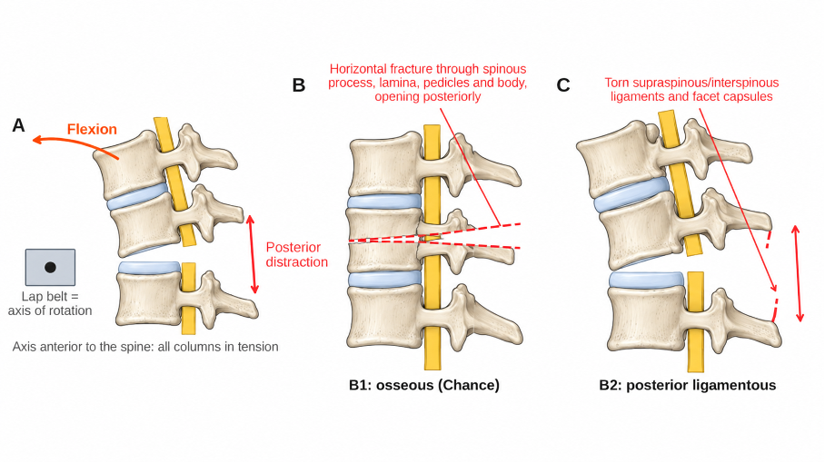
Figure 1. Flexion-distraction injuries. (A) Mechanism: flexion around an axis located anterior to the spine (lap belt) produces tension failure of the posterior and middle columns. (B) Osseous B1 (Chance) pattern with a horizontal fracture through the posterior elements and vertebral body, opening posteriorly. (C) B2 pattern with disruption of the posterior ligaments and facet capsules.
Treatment
Purely osseous B1 injuries in neurologically intact patients have good healing potential. They can be treated with reduction in extension and a hyperextension cast or thoracolumbosacral orthosis, especially in children and adolescents. Upright radiographs in the cast or brace, repeated during the first weeks, detect loss of reduction. Immobilization usually continues for about 3 months until healing is confirmed.
The most common error is to treat a B2 injury as a stable compression or burst fracture; when posterior ligamentous integrity is uncertain, MRI should be obtained before choosing nonoperative care.
Ligamentous and osseoligamentous B2 injuries heal poorly and are treated surgically, because nonoperative care risks progressive kyphosis and chronic pain [3]. The TL AOSIS mirrors this approach: B1 injuries score 5 points (equivocal), whereas B2 injuries score 6 (operative) [6]. Posterior instrumentation in compression restores the tension band; short constructs, one level above and below, are usually sufficient when the anterior column is intact. Overcompression should be avoided when the middle column is comminuted or a disc herniation is present, as it may push material into the canal. Percutaneous fixation is a reasonable option for B1 injuries in adults, including patients with abdominal injuries who cannot tolerate a cast. Abdominal injuries take priority, and spinal fixation is timed with the general surgical plan. Prone positioning on a frame often reduces the deformity. Fluoroscopy confirms restoration of posterior height before final tightening, and a disc herniation identified on MRI is removed before compression is applied.