Figures
Overview
OVCFs are particularly common in elderly postmenopausal women and in patients receiving long-term glucocorticoids. They occur most often at the thoracolumbar junction (T10–L2), followed by the mid-thoracic spine.
Clinical Presentation
In patients who present clinically, sudden back pain is the most common symptom. It usually follows minor trauma, but some patients recall no injury. Pain increases with movement and decreases with rest. Local tenderness and paraspinal muscle spasm at the fracture level are common.
Multiple fractures cause kyphotic posture, height loss and functional limitation. Neurological deficit is rare and is usually associated with posterior wall involvement.
Imaging
Lateral and anteroposterior radiographs are the first step. Standing views show the true degree of collapse, and comparison with supine images reveals instability or an intravertebral cleft. CT assesses the integrity of the posterior wall and the fracture lines.
MRI is the most sensitive method for acute fractures. STIR sequences show bone marrow edema and distinguish new from old (chronic) fractures, which helps identify the painful level. Bone densitometry and laboratory tests for secondary osteoporosis complete the evaluation.
Classification
The OF classification of the German Society for Orthopaedics and Trauma (DGOU) describes fracture morphology and stability in five types, from OF 1 (edema only) to OF 5 (tension-band injury) (Figure 1) [1]. The extent of posterior wall involvement (none or minor, <1/5, versus distinct, >1/5) separates OF 2 from OF 3, and OF 4 and OF 5 fractures are generally unstable.
The OF score adds clinical modifiers (bone density, pain under analgesia, fracture progression, neurological status, mobility and general health) and guides the choice between nonoperative and operative treatment [2].
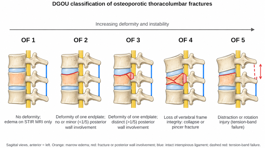
Figure 1. DGOU classification of osteoporotic thoracolumbar fractures. OF 1, no deformity, with marrow edema on STIR MRI only; OF 2, deformity of one endplate with no or minor (<1/5) posterior wall involvement; OF 3, deformity of one endplate with distinct (>1/5) posterior wall involvement; OF 4, loss of vertebral frame integrity, vertebral body collapse or pincer-type fracture; OF 5, distraction or rotation injury with tension-band failure.
Treatment
Nonoperative treatment is appropriate for most stable fractures. It includes analgesics, a short period of rest followed by early mobilization, physiotherapy and, when needed, a brace or corset for mechanical support. Prolonged bed rest should be avoided.
Medical treatment of osteoporosis is essential: calcium 1000–1200 mg/day, vitamin D 800–1000 IU/day and pharmacological therapy. A recent vertebral fracture or multiple vertebral fractures indicate very high risk; an anabolic agent (teriparatide, abaloparatide, romosozumab) followed by an antiresorptive is recommended as first-line therapy where available, and a bisphosphonate or denosumab is used otherwise [3].
Vertebroplasty or kyphoplasty may be considered when pain persists despite 4–6 weeks of conservative treatment or when collapse progresses. Kyphoplasty partly restores vertebral height and has a lower risk of cement leakage. Evidence is mixed: kyphoplasty was superior to nonsurgical care in a randomized open-label trial [4], whereas vertebroplasty was not superior to a sham procedure [5]. Augmentation is therefore reserved for carefully selected patients [6].
Surgical stabilization is needed in a minority of patients, mainly those with unstable OF 4 and OF 5 fractures. Posterior instrumentation is combined with cement-augmented screws, longer constructs or augmentation of the fractured vertebra to improve fixation in osteoporotic bone [2].
Surgical Indications
• Unstable fractures (OF 4 and OF 5)
• Neurological deficit
• Progressive collapse or kyphosis
• Persistent severe pain despite optimal nonoperative treatment (augmentation in selected patients)
Prognosis
With appropriate treatment, most patients achieve pain control and early mobilization. However, kyphosis may progress and reduce pulmonary function. About one in five women sustains another vertebral fracture within a year [7]; this imminent risk is a reason to start treatment promptly. Long-term outcome depends on adherence to antiosteoporotic treatment and rehabilitation.
Complications
• Augmentation-related: cement leakage, pulmonary cement embolism, infection
• Late: Kümmell disease (delayed collapse due to osteonecrosis), new fractures at adjacent levels
• Surgery-related: screw loosening, implant failure, proximal junctional failure
Careful patient selection, sound technique and medical treatment of bone health reduce these risks.
Differential Diagnosis
• Pathological fracture due to metastasis or myeloma (convex posterior wall, pedicle involvement, soft-tissue mass, diffuse marrow replacement)
• Spondylodiscitis
• High-energy traumatic fracture
• Scheuermann disease and developmental wedging
References
1. Schnake KJ, Blattert TR, Hahn P, et al. Classification of osteoporotic thoracolumbar spine fractures: recommendations of the Spine Section of the German Society for Orthopaedics and Trauma (DGOU). Global Spine J. 2018;8(2 Suppl):46S-49S.
2. Blattert TR, Schnake KJ, Gonschorek O, et al. Nonsurgical and surgical management of osteoporotic vertebral body fractures: recommendations of the Spine Section of the German Society for Orthopaedics and Trauma (DGOU). Global Spine J. 2018;8(2 Suppl):50S-55S.
3. Kanis JA, Harvey NC, McCloskey E, et al. Algorithm for the management of patients at low, high and very high risk of osteoporotic fractures. Osteoporos Int. 2020;31(1):1-12. doi:10.1007/s00198-019-05176-3
4. Wardlaw D, Cummings SR, Van Meirhaeghe J, et al. Efficacy and safety of balloon kyphoplasty compared with non-surgical care for vertebral compression fracture (FREE): a randomised controlled trial. Lancet. 2009;373(9668):1016-24. doi:10.1016/S0140-6736(09)60010-6
5. Firanescu CE, de Vries J, Lodder P, et al. Vertebroplasty versus sham procedure for painful acute osteoporotic vertebral compression fractures (VERTOS IV): randomised sham controlled clinical trial. BMJ. 2018;361:k1551. doi:10.1136/bmj.k1551
6. Ebeling PR, Akesson K, Bauer DC, et al. The efficacy and safety of vertebral augmentation: a second ASBMR task force report. J Bone Miner Res. 2019;34(1):3-21. doi:10.1002/jbmr.3653
7. Lindsay R, Silverman SL, Cooper C, et al. Risk of new vertebral fracture in the year following a fracture. JAMA. 2001;285(3):320-3. doi:10.1001/jama.285.3.320