Bone Transport and the surgical management of bone defects.
Overview
Definition: A specialized surgical technique used to reconstruct critical-sized bone defects resulting from high-energy trauma, infection (osteomyelitis), or tumor resection.
Mechanism: Utilizes the body’s natural ability to regenerate tissue when subjected to controlled mechanical tension, a process known as distraction osteogenesis (DO).
Clinical Goal: To reconstitute bone stock and obtain a stable, functional limb as an alternative to amputation or complex bone grafting.
Biological Foundations
Distraction Osteogenesis: The core principle involving the gradual translocation of a healthy bone segment across a gap.
Vascularity: Successful transport requires a biologically sound healing environment. Well-vascularized bone and competent soft-tissue coverage are essential for producing healthy "regenerate" (new bone).
Evaluation: Tools such as MRI and arteriography are utilized to assess the vascular health of the marrow and surrounding tissues prior to surgery.
Treatment Strategies & Planning
The choice of procedure is dictated by the size of the defect and vascular status:
Acute Shortening:
Indicated for defects up to 3–4 cm in the tibia and 5–7 cm in the femur.
Risk: Excessive shortening can cause vascular "kinking" or tissue necrosis.
Gradual Shortening: For larger defects, shortening at a rate of 0.5 cm per day helps avoid soft-tissue and vascular complications.
Bone Transport: Typically indicated for defects greater than 3–4 cm. Massive defects (>10–12 cm) often require combination methodologies to reduce treatment time.
Clinical Phases of Bone Transport
Corticotomy: A low-energy surgical cut is made in healthy bone while preserving the periosteum and blood supply.
Latency Phase: A waiting period of 5 to 14 days (or 7-10 days depending on protocol) allows the initial healing response to begin before movement.
Transport Phase: The segment is moved toward the defect at a rate of approximately 1 mm per day, often divided into smaller increments (e.g., 0.25 mm four times daily).
Docking: The point where the transported segment reaches the target bone.
Consolidation Phase: The frame remains in place while the new "regenerate" matures and develops a neocortex until it can support weight-bearing.
Surgical Approaches & Techniques
Bifocal Approach: Involves two active segments; one site undergoes compression (closing the defect) while another undergoes distraction (lengthening).
Trifocal Approach: Utilizes three active segments (one compression site and two lengthening sites); preferred for large defects to reduce total treatment time.
Implementation Methods:
All-External: Circular (Ilizarov) or Taylor Spatial Frames (TSF). TSF allows for precise adjustment of angulation and translation.
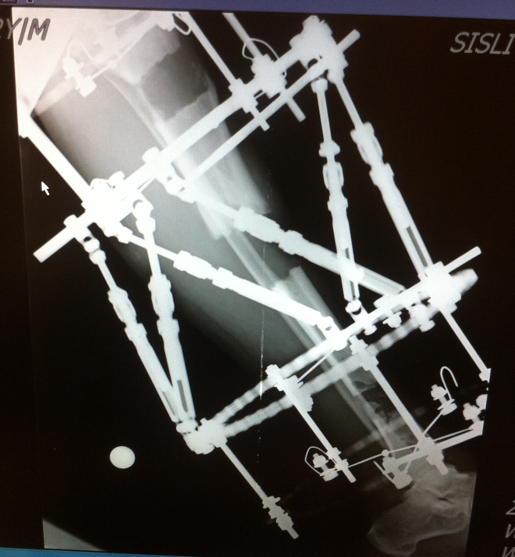
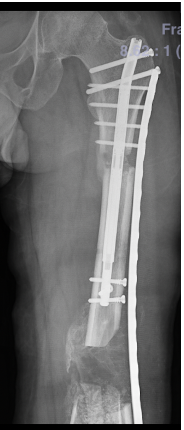
Hybrid Techniques: Combines external fixators with internal implants (e.g., Bone Transport Over a Nail - BTON) to shorten the External Fixation Time (EFT).
All-Internal: Uses motorized telescopic intramedullary nails or Plate-Assisted Bone Segment Transport (PABST) to improve patient comfort.
Management of the Docking Site
Once segments meet, the docking site often requires surgical intervention.
Procedures: Typically involves removing scar tissue, freshening bone ends, and applying bone grafts to ensure a solid union.
Complications & Clinical Significance
Common Complications:
Nonunion at the docking site (most frequent).
Pin tract infections and joint stiffness (common with external fixators).
Inadequate regenerate leading to bone collapse or deformity.
Clinical Significance: Considered a cornerstone of limb salvage, especially for infected non-unions, as vascularized new bone formation helps eradicate infection.
Future Directions: Increasing use of orthobiologics (growth factors) and internal lengthening nails to reduce the time spent in external fixation.