LIMB LENGTHENING
I. Introduction and Definition
Limb-Length Discrepancy (LLD): Defined as a significant difference between the lengths of lower or upper limbs.
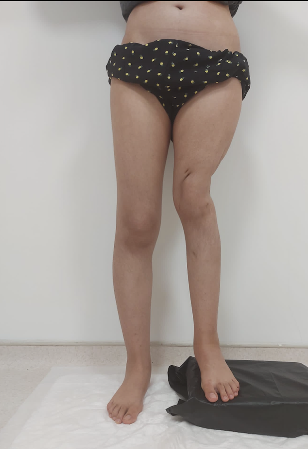
Etiology:
Congenital: e.g., fibular hemimelia.
Developmental: e.g., growth arrest or childhood illness.
Acquired: e.g., post-traumatic malunion or infection.
Primary Goal: Achieve functional leg-length equality by skeletal maturity to improve mobility and prevent long-term complications like compensatory scoliosis and back pain.
Distraction Osteogenesis (DO): Also known as "callotasis," it uses controlled microtrauma (osteotomy) to trigger the body's innate regenerative capacity to grow new bone in a precise direction.
II. Historical Evolution
Early 20th Century: Alessandro Codivilla first applied skeletal traction for bone lengthening.
Mid 20th Century: Leroy Abbott introduced slow continuous traction to overcome soft tissue resistance.
1950s (The Breakthrough): Gavril Ilizarov discovered the "law of tension-stress," proving that gradual traction on living tissues stimulates active growth and regeneration.
1970s: Wagner method used unilateral fixators but faced high complication rates.
III. Evaluation and Growth Prediction
Clinical Evaluation:
Graduated blocks: Used to level the pelvis while standing to determine functional discrepancy.
Tape measurements: From ASIS to medial malleolus to distinguish "true" (skeletal) from "apparent" (pelvic obliquity) LLD.
Radiological Assessment:
Teleoroentgenogram: The gold standard; a full-length standing radiograph.
Must account for the pelvis and hindfoot to avoid underestimating discrepancy.
Growth Prediction (Children):
Multiplier Method: Regarded as the most accurate; uses age-specific multipliers.
Other Methods: Moseley straight-line graph and Menelaus arithmetic method.
IV. Clinical Indications
Limb Length Discrepancy: Typically those exceeding 3 cm.
Congenital Deficiencies: e.g., Hip or skeletal dysplasias.
Large Bone Defects: Reconstructing bone lost to trauma, chronic infection (nonunion), or tumor resection.
Other Applications:
Limb Salvage: Treating Stage 4 diabetic foot ulcers to prevent amputation.
Achondroplasia: For patients with dwarfism.
Hand Reconstruction: Lengthening digits (thumbs) to restore function.
Cosmetic Lengthening: Available for short stature but requires mandatory psychiatric evaluation for body dysmorphic disorder.
V. Biological Principles and Molecular Mechanisms
The Five Biological Phases:
Osteotomy: Surgical cut, typically in the metaphysis due to dense blood supply.
Distractor Application: Stability via external (Ilizarov) or internal (intramedullary) devices.
Latency Phase: A waiting period of 5–10 days (5–7 for femur, 10–14 for tibia) allowing hematoma formation.
Distraction Phase: Bone segments are pulled apart at 1 mm per day.
Rhythm: Usually split into four 0.25 mm increments to optimize bone quality.
Consolidation Phase: The device stays in place for 4–6 months as "regenerate" bone matures and mineralizes.
Molecular Drivers:
Pro-inflammatory Cytokines: IL-1, IL-6, and TNF-α recruit mesenchymal stem cells early on.
Growth Factors: BMPs (Bone Morphogenetic Proteins), VEGF (for blood vessels), and TGF-β stimulate osteoblast activity.
Mechanical Tension: Maintains high levels of IL-6, which temporarily slows bone maturation to allow continued lengthening.
VI. Surgical Strategies and Fixation Evolution
Epiphysiodesis: A simpler procedure that arrests growth in the longer limb's physis during puberty.
Fixation Methods:
External Fixators: Ilizarov apparatus or Hexapod systems (e.g., TSF) which use computer-assisted technology for multi-plane correction.
Lengthening Over Nail (LON): Combination of external and internal fixation.
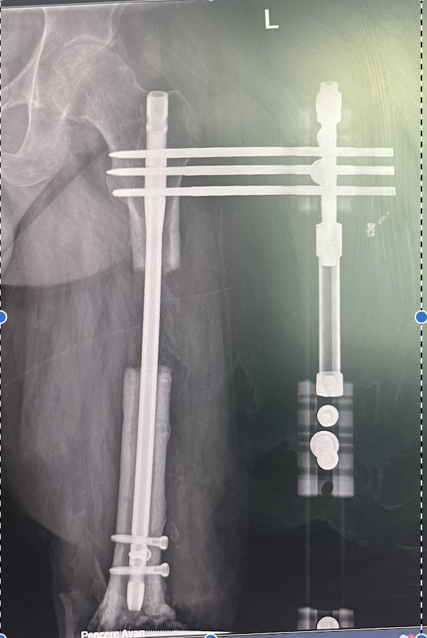
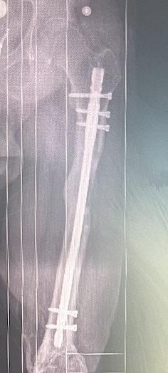
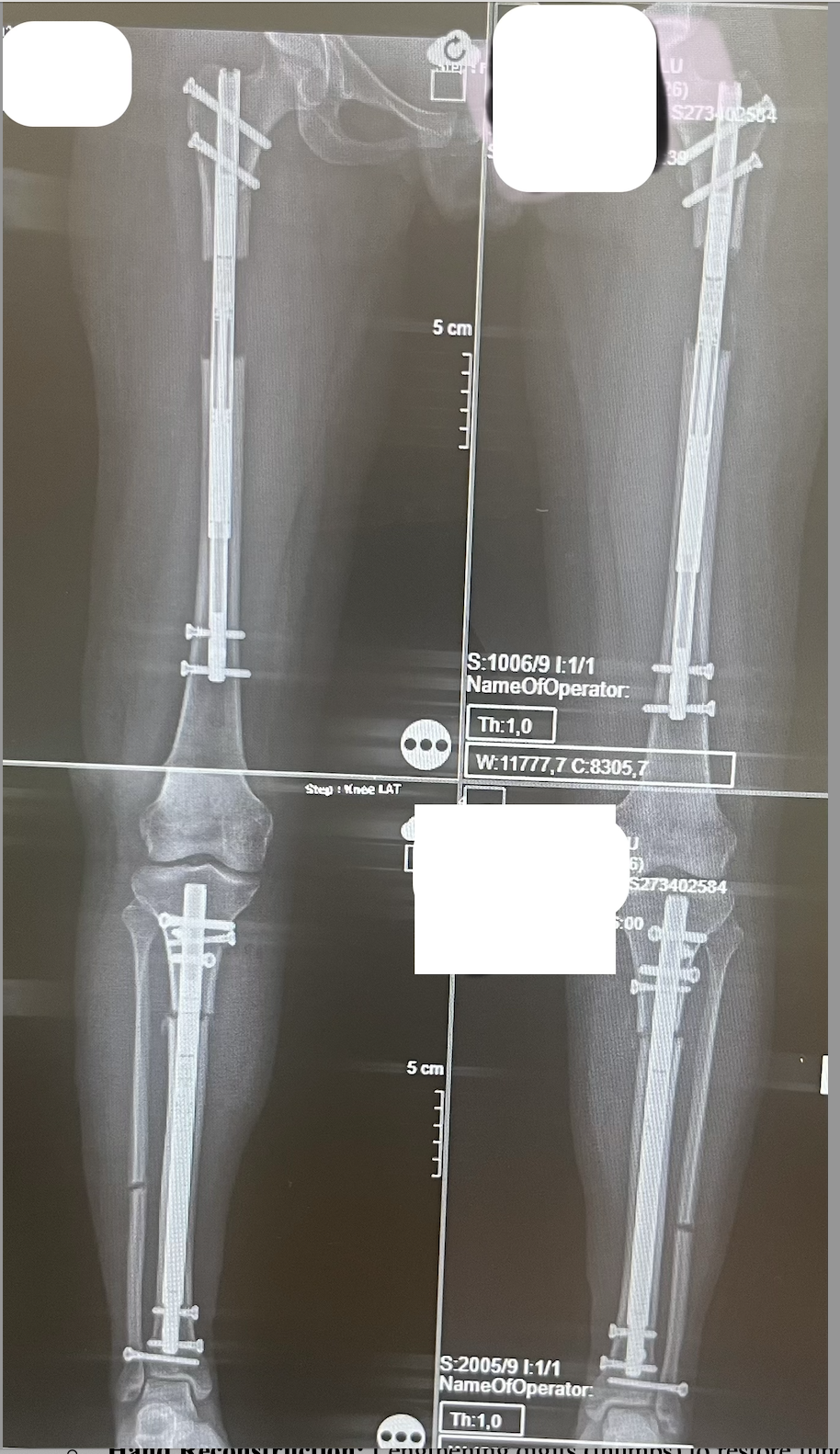
Internal Motorized Nails:Fully implantable devices like Fitbone or PRECICE (magnetic) that eliminate external hardware and reduce infection risk
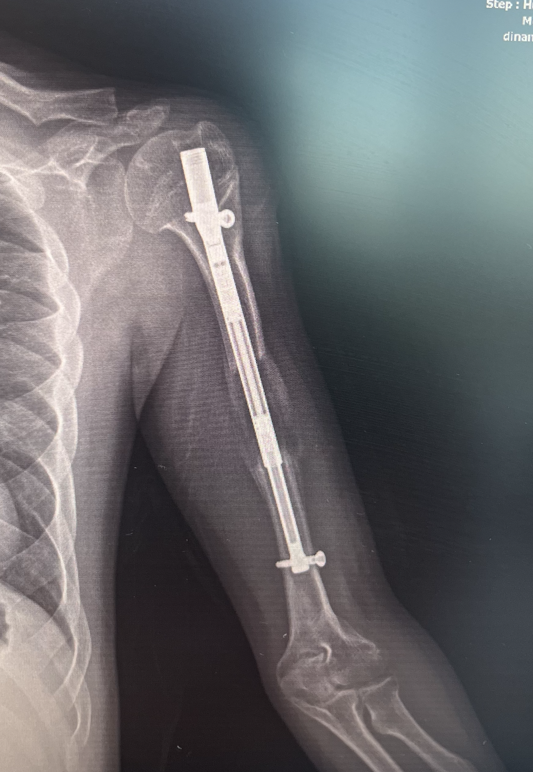
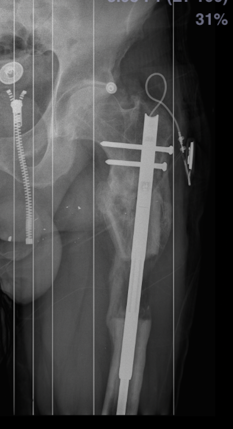
Correction Order: Angulation and length should generally be corrected simultaneously, followed by rotation and translation.
VII. Step-by-Step Intramedullary Procedure
Surgical Venting: "Vent holes" are drilled to allow marrow fat to escape, preventing fat embolism.
Periosteal Elevation: Careful preservation of the periosteum is essential for blood supply.
Nail Insertion: Percutaneous osteotomy followed by immediate nail advancement across the site.
Securing Stability: Use of transverse locking screws and blocking screws to prevent fragments from tilting in wide bone regions.
Distraction: Controlled by an External Remote Controller (ERC) interacting with the internal magnet.
Rehabilitation: Intensive physiotherapy is vital to prevent joint contractures; functional loading (partial weight-bearing) stimulates bone maturation.
Implant Removal: Nail is typically removed 1 to 2 years after consolidation.
VIII. Risks, Complications, and Management
Technical Risks:
Pin Track Infections: Most common issue with external fixators.
Nerve Damage: Peripheral neuropathy; the common fibular nerve is highly vulnerable during tibial lengthening.
Joint Stiffness: Contractures due to soft tissues struggling to adapt.
Bone Issues: Poor regeneration, axial malalignment, or fractures if the new bone is fragile (osteopenia).
Patient Factors: Success is lower in patients who are smokers (nicotine users), diabetic, or malnourished.
Psychosocial Burden: Bulky hardware and long treatment duration (up to 18 months) can cause significant psychological distress.
IX. Future Directions and Innovations
Internal Hardware: Continued shift toward motorized/magnetic nails to reduce infection and psychosocial impact.
Biological Enhancements: Use of platelet-rich fibrin (PRF), BMP injections, or hyperbaric oxygen to accelerate bone maturation.
Customization: Development of 3D-printed custom distractors and biodegradable materials for growth factor release.